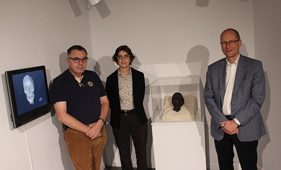
The HÂş» Art Gallery, located in the basement of the HÂş» Arts Centre, is currently playing host to a much older guest than it is usually accustomed to. Since mid-September, the mummified head of an ancient Egyptian has been on display in an exhibit entitled From the Vault: The Oldest Patient.
The head itself was donated to the gallery in 1974 by David Donald Cameron Mackay. Mackay, former president of the Nova Scotia College of Art, was fond of collecting objects of aesthetic and historic significance. How it came into Mackayâs possession, like so much else about the mummy, remains a mystery. Â
A new (old) patient
According to Michele Gallant, registrar and preparator of the gallery and its permanent collection, while HÂş» has had the head in its possession for the last four decades, it has never been publicly displayed or closely examined; even its age is unknown.
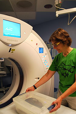
 It is precisely this mystery that the exhibit attempted to solve. The Oldest Patient is a collaboration between the gallery and two faculty members of HÂş» Medical School. Gallant began her investigation by contacting Dr. William Baldridge, head of the Department of Medical Neuroscience at HÂş», who pointed her in the direction of two radiologists with very similar names: Dr. Matthias H. Schmidt and Dr. Pierre Schmit. The duo agreed with interest to perform a CT scan on the head and apply their medical knowledge in an attempt to uncover new details about their patient.
Both Gallant and Dr. Schmit intentionally use the term âpatientâ when discussing the head to convey a sense of humanity.
âThese are human remains we are speaking about,â says Gallant, ânot an object, or a relic, but remains.â Dr. Schmidt adds that, âputting [Patient] in the title was an indication of respect.â
When one looks at the history of western fascination with mummified remains, the reasoning behind this becomes quite clear. Prior to the widespread use of diagnostic technology it was common for museums to unveil linen-wrapped mummies, often publicly, in order to examine what lay underneath. This practice, aside from being insensitive to the remains of the deceased, can also be quite damaging to the physical integrity of the body.
Exploring beneath the skin
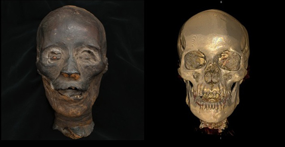
While the head currently on display at HÂş» Art Gallery is not wrapped in linen, and thus has a fully visible face, it was what lies underneath the face that interested the team. Through the use of CT imaging they were able to get a look at the internal structure of their patientâs head.

Dr. Schmit explains the findings are concurrent with what is known about the mummification process: the patientâs nasal wall had been pierced, and the brain removed, replaced with a resinous substance. What may be more interesting, however, are the smaller findings that shed light on the way the patient lived. The patientâs teeth, for instance, were worn down, with many cavities. Dr. Schmit posits this is most likely due to the methods of grinding flour at the time, in which sandstone was used, often contaminating the finished product with particles of sand that would wear down the enamel.
There were questions the team was not able to answer, though â such as the nature of the resinous substance inside the patientâs cranium, or the exact age of the head â because they would require more intrusive tests which might compromise the integrity of the mummy.
At the end of the day Gallant considers the project and its findings to be a success, calling it a âwonderful collaboration with the Med School and the Department of Radiology.â
The exhibit will be on display until this coming Sunday, November 22 in the .


