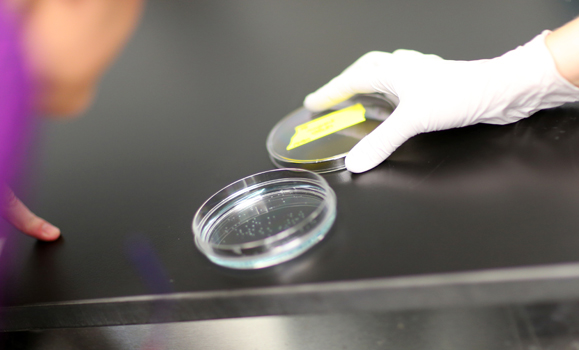
For those with a mind for science, like Victoria Bentley, a lab is the hotspot of discovery.
A HÂţ» student since 2012, Bentley — formerly a masters student in the Department of Pathology who’s now finishing the first year of her MD degree — is a co-author of two cancer studies that have bubbled up from HÂţ» Medical School’s Zebrafish Core Facility.
The two studies use a technique called xenotransplantation. Pioneered by Drs. Graham Dellaire and Jason Berman, associate professors in the Departments of Pathology and Pediatrics, respectively, the technique was used to transplant human leukemia cells into zebrafish embryos, allowing Bentley to study their growth and test anti-cancer therapies.
A rare form of leukemia
The group’s research focuses on a specific type of leukemia — a cancer of the white blood cells — called T-cell acute lymphocytic leukemia (T-ALL).
Under normal conditions, blood stem cells in our body become one of two types of basic cells: red blood cells, and white blood cells, which help fight infection in the body. But when blood stem cells in the bone marrow grow and behave outside of their normal parameters, they develop into leukemia cells. These cells are the cause of the cancer the co-authors are studying.
According to the studies’ authors, there are about 300 cases of childhood acute leukemia in Canada each year. 80 per cent of those are acute lymphoblastic leukemia (ALL). T-ALL specifically targets our body’s T-cells and accounts for approximately 15 per cent of ALL diagnoses.
“It’s a rare, high-risk subset of leukemia,” says Bentley.
The first study, published in Haematologica, demonstrates for the first time that T-cell leukemia cells from a patient’s bone marrow biopsy can be successfully xenografted in zebrafish.
The process involves microinjecting fluorescently labeled patient tumor cells into the yolk sac of a 48-hour-old zebrafish embryo (shown in the photo below). As the zebrafish continues its natural growth, the T-ALL cells engraft and grow in the fish. This allows treatment response to be tested in the lab and provides information on the best therapy for a specific patient.

The therapies the group examined specifically target genetic mutations commonly found in T-ALL and inhibit hyper activation of growth signals and cell proliferation. An effective drug is identified when the xenografted cells in the zebrafish no longer divide — or ultimately die — compared to cells in fish without a drug treatment.
Eventually, the research could lead to better personalization of therapy for T-cell leukemia patients.
“Our zebrafish xenograft model allowed us to determine the susceptibility of a patient’s T-ALL within three days,” says Dr. Dellaire. “If we were using this information to guide therapy, we could rapidly identify an effective drug and personalize a patient’s therapy, reducing side-effects and improving outcome.”
Protecting the heart
The co-authors are also using a zebrafish model to test protectants for cardiotoxicity: the damaging effects of chemotherapy on the heart.
Published in Science Translational Medicine, the group’s second high-impact study examined the chemotherapy drug doxorubicin, a common treatment for leukemias and many adult cancers, including breast cancer.
Although widely used, children treated with doxorubicin for leukemia often suffer heart damage, predisposing them for heart failure decades after successful treatment. The drug can cause similar cardiac damage to adult patients, increasing the chance of post-therapy heart failure.
Dr. Berman, chair of the Zebrafish Core Facility and one of its main tenants, says his studies on chemoprotectants began through a collaboration with Dr. Randall Peterson at Massachusetts General Hospital and Harvard Medical Centre.
“When zebrafish embryos are treated in water with doxorubicin, they develop abnormalities of the heart,” explains Dr. Berman. “Dr. Peterson identified a number of compounds that protected the zebrafish heart when exposed to this drug. One of the most promising compounds was a drug called visnagin.”
The data from Dr. Peterson’s work was applied to Drs. Berman and Dellaire’s human leukemia zebrafish transplant research. When paired with visnagin, the doxorubicin treatment effectively killed the human leukemia cells xenografted in zebrafish but protected the zebrafish heart from doxorubicin-induced heart damage.
“It’s incredibly exciting to think that we could customize a patient’s treatment strategy with the hope of minimizing toxicities and improving outcomes,” says Bentley.
Student-led research at HÂţ»
“I have a longstanding love for cancer research,” says Bentley. As the lead on the zebrafish studies, Bentley designed and performed experiments, analyzed data, wrote the papers and presented the research at conferences.
“Being able to do clinically relevant research using a zebrafish xenotransplantation model is exciting, and this new model presents a number of opportunities. I was able to evaluate the sensitivity of T-ALL cell lines and patient samples from children, all in a clinically-actionable timeframe.”
Bentley says she hopes the research will improve treatments for patients with leukemia and improve the safety of widely-used therapies like doxorubicin. And while these studies focus on childhood leukemia, she says the research is an important step towards better, less harmful therapies for patients being treated for a wide range of cancers.
“This research provides a compelling rationale for the use of the zebrafish xenotransplantation model. This is impossible just using cell-based approaches, and is much faster than mouse models.”
As a medical student at HÂţ», Bentley will be continuing with the studies through the school’s Research in Medicine (RIM) program.
“I’ll be back in the lab during the summer as part of the RIM program, using the zebrafish to look at novel therapeutics for T-ALL. I feel very fortunate to have this opportunity and am looking forward to a busy and rewarding summer.”

